

Liver nodule: is it always cancer?
A liver nodule is not always cancer. Learn the most common benign lesions, how a liver nodule is investigated, and when it needs monitoring.
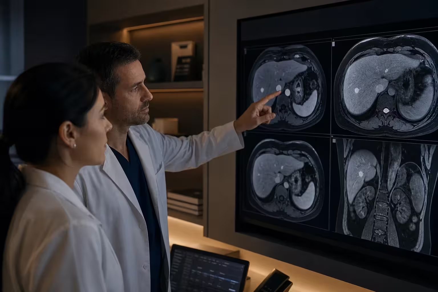
Being told about a liver nodule usually causes alarm and an immediate worry about cancer. This is an understandable reaction, but it is important to clarify from the outset: a liver nodule is not always cancer. In fact, most liver lesions found by chance are benign and pose no threat to life. Even so, each case needs to be assessed carefully, because some lesions warrant more detailed investigation. This text gathers clear and responsible information to help you understand what a hepatic nodule is, what the most common causes are, how it is investigated, and in which situations monitoring or treatment is needed — without ever replacing an individual assessment by a physician.
What a liver nodule is
The term liver nodule describes an area of the organ that appears different from the surrounding tissue on an imaging study. Also called a hepatic nodule or liver lesion, it can have many different origins: it may be a cluster of blood vessels, a collection of fluid, a benign growth of cells, or, in a minority of cases, a lesion that requires investigation to rule out malignancy. Simply finding a nodule, on its own, does not define the diagnosis — it is the combination of the lesion's features and the patient's context that guides the approach.
A liver nodule is not always cancer
It is worth repeating, because this is the central concern of anyone who receives the result: the vast majority of liver nodules are benign. Many are incidental findings, that is, discovered by chance during tests done for another reason, in people without any symptoms. Benign lesions such as hemangiomas and cysts are quite common in the general population. This does not mean that every nodule can be ignored, but rather that finding a lesion should not be interpreted, right away, as synonymous with cancer. The role of the medical assessment is precisely to distinguish, calmly and methodically, what is benign from what warrants additional attention.
The most common benign lesions
There are several benign liver lesions, each with its own behavior and features. Knowing about them helps explain why many nodules do not require treatment, only diagnostic confirmation and, in some cases, monitoring.
Hemangioma
The hemangioma is one of the most common benign lesions of the liver. It is a tangle of blood vessels, usually without any symptoms and discovered by chance. Most of the time, it remains stable over time and does not turn into cancer. When the imaging features are typical, the diagnosis is usually reassuring and often requires no intervention — only confirmation and, occasionally, observation.
Focal nodular hyperplasia
Focal nodular hyperplasia is another common benign lesion, made up of liver cells organized in a particular way. It is usually asymptomatic and, in general, carries no risk of becoming malignant. Recognizing it with specific imaging studies helps avoid unnecessary procedures, since, when well characterized, the usual approach is conservative.
Hepatic adenoma
Hepatic adenoma is a benign growth of liver cells, less common than the lesions above. Although it is benign, in certain situations it may warrant special attention, depending on the size, the features, and the clinical context. For this reason, when an adenoma is suspected, specialized assessment is particularly important to define the best approach, which may range from monitoring to interventions in selected cases.
Hepatic cysts
Cysts are collections of fluid within the liver, very common and, most of the time, entirely benign. Simple cysts are usually harmless and require no treatment. In rarer situations, cysts with different features may need additional investigation. Ultrasound usually distinguishes a simple cyst from other lesions well.
When a nodule may be malignant
Although most are benign, a portion of liver nodules may correspond to malignant lesions. These include tumors that originate in the liver itself and also metastases, that is, lesions that reach the liver from tumors located in other organs. The risk that a nodule is malignant varies with the context: factors such as prior chronic liver disease, a history of cancer in another organ, and certain imaging features may increase this concern. Precisely for this reason, the assessment considers not only the nodule itself but the person's entire clinical history.
It is important to stress that the possibility of malignancy is not the rule, but a hypothesis to be confirmed or ruled out through an organized investigation. The goal is not to alarm, but to ensure that no lesion requiring care goes unnoticed.
How nodules are usually discovered
Most liver nodules are discovered incidentally, during tests performed for other reasons — such as an abdominal ultrasound ordered to evaluate pain, gallstones, or as part of a check-up. Because many of these lesions cause no symptoms, the person often learns of the nodule's existence without ever having felt anything. In some cases, symptoms such as abdominal discomfort may prompt testing, but this is less common than an incidental finding.
How the nodule is characterized
Once identified, the next step is to characterize the nodule, that is, to understand its features in order to define its nature. Ultrasound is usually the first test, as it is widely available, painless, and without radiation. When needed, the physician may request more detailed studies for an in-depth analysis:
- Computed tomography, which provides detailed images and helps assess the pattern of the lesion.
- Magnetic resonance imaging, especially useful for characterizing liver lesions in great detail.
- Laboratory tests, which help assess liver function and the clinical context.
- Biopsy, in selected situations, when imaging studies are not enough to clarify the nature of the nodule.
In most cases, the combination of clinical history and imaging studies allows the lesion to be characterized with good accuracy, without the need for a biopsy. The choice of tests depends on each situation and is decided by the responsible physician.
Benign versus concerning features
Imaging studies help identify patterns that guide the interpretation of the nodule. Some features tend to be reassuring, while others indicate the need for deeper investigation. In general, lesions with a typical appearance of known benign conditions, and that remain stable over time, tend to be considered more reassuring. Lesions with atypical features, progressive growth, or that arise in a liver with prior disease, on the other hand, usually warrant additional attention.
It is essential to understand that this analysis is technical and must be carried out by qualified professionals. Trying to interpret a report on your own, without the proper context, can cause unnecessary distress or, conversely, false reassurance. Each finding has meaning only within the patient's complete picture.
Why comparing with previous tests helps
One of the most valuable resources in assessing a liver nodule is time. When old imaging studies are available, comparing the current images with the previous ones makes it possible to see whether the lesion has changed in size or appearance over the months or years. A lesion that remains stable over a prolonged period is usually a reassuring sign, while more evident changes may indicate the need for additional investigation. This sense of evolution over time is information that a single isolated test cannot provide.
For this reason, whenever possible, it is important to gather and bring to the consultation tests performed in the past, even if they were done for other reasons. These older images can save steps, avoid unnecessary procedures, and give the physician a more complete overview. Keeping your reports and images organized, including digital files when available, is a simple habit that can make a difference in the assessment of a hepatic nodule.
When to simply monitor and when to investigate or treat
One of the most frequent questions is whether the nodule needs treatment. The answer depends on the nature of the lesion. Many typical benign lesions, such as hemangiomas and simple cysts, do not require treatment and, in several cases, do not even need ongoing monitoring once well characterized. Other benign lesions may be monitored with periodic imaging to confirm that they remain stable. When there is doubt about the nature of the nodule or a suspicion of malignancy, however, the investigation is deepened and the approach is defined case by case.
Monitoring, when indicated, usually involves repeating imaging studies at intervals determined by the physician. This strategy makes it possible to observe the behavior of the lesion over time, which is valuable information for confirming benignity or identifying any change that warrants attention.
The role of the specialist
Assessing a hepatic nodule benefits from the experience of a professional accustomed to dealing with liver diseases, such as a digestive system and liver surgeon or a hepatologist. This specialized perspective helps integrate the information from the tests with the clinical history, define whether and when complementary tests are needed, and avoid both unnecessary interventions and delays in care. The goal is to offer a balanced approach: neither excessive in the face of a clearly benign lesion, nor negligent toward one that requires attention.
When surgery is considered
For most liver nodules, especially benign and asymptomatic ones, surgery is not necessary. It is usually considered in specific situations — for example, when there is a suspicion of malignancy, when the lesion has features that increase risk, when it causes significant symptoms, or when there is a diagnostic doubt that cannot be resolved by other means. Even in these cases, the decision is always individualized and made together with the patient, after a careful assessment of risks and benefits.
When indicated, the surgical approach to the liver is planned carefully, considering the location and features of the lesion. The key point is that surgery is not the automatic answer for every nodule, but an option reserved for well-defined situations.
How to cope with waiting for results
The interval between finding a liver nodule and completing the investigation can be distressing. It is natural to feel worried, but it is worth remembering that, in most cases, there is time to assess the lesion calmly and methodically, without rushed decisions. Haste rarely improves the outcome; organization and appropriate follow-up do. Understanding that the investigation follows steps helps you get through this period with greater peace of mind.
Some attitudes help during this time: following the physician's guidance about the tests, avoiding self-diagnosis based on internet searches, writing down questions to clarify at the appointment, and seeking support from people you trust. If anxiety is very intense, telling the professional is also valid — they can offer explanations that help reduce fear and better understand each stage of the assessment.
Frequently asked questions about liver nodules
Is a liver nodule dangerous?
Most of the time, no. A large share of liver nodules are benign and pose no risk. What defines whether a lesion is dangerous is the combination of its features and the clinical context, assessed with appropriate tests. That is why correct characterization is so important.
Can a liver nodule turn into cancer?
Most benign lesions, such as hemangiomas, focal nodular hyperplasia, and simple cysts, do not turn into cancer. Some specific lesions may require special attention under certain circumstances. Individual assessment is what makes it possible to understand the real risk in each case.
Do I need to repeat tests if the nodule is benign?
It depends on the type of lesion. Some typical benign lesions, once well characterized, do not require ongoing monitoring. Others may be monitored with periodic tests to confirm stability. The physician defines the need and frequency according to each situation.
Does a liver nodule cause symptoms?
Many nodules cause no symptoms and are discovered by chance. In some cases, larger lesions may cause abdominal discomfort. The absence of symptoms, however, does not rule out the need to characterize the lesion properly.
When to see a specialist
If a test has revealed a liver nodule, the best course is to take the result to a physician experienced in liver diseases for a proper assessment. Avoid drawing hasty conclusions from an isolated report or from internet searches, which can cause unnecessary anxiety. A specialized consultation makes it possible to understand the nature of the lesion, decide whether any complementary test is needed, and establish, when appropriate, a monitoring plan. Bringing your existing tests, including previous images if available, and a list of questions helps make the assessment more complete and objective.
Understanding that a liver nodule is not always cancer is the first step toward dealing with the finding more calmly. In most cases, there is time to investigate carefully, clarify the nature of the lesion, and decide, together with a trusted professional, whether it simply needs to be observed or warrants some additional approach — avoiding both unnecessary fear and a lack of care when it is truly indicated.
Do you have questions about your case?
A specialized evaluation helps understand the best path, with clarity and security.